2026 Best Dental Technologies for the Future of Dentistry

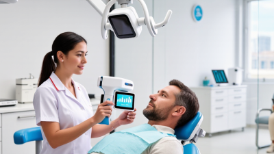
The future of dentistry is already taking shape in ordinary clinical moments: a scanner replacing impression trays, a monitor displaying a tooth in three dimensions, and software helping clinicians compare treatment options. These tools can improve precision and communication, but they do not make sound judgment automatic. A detailed digital image still needs an experienced clinician to interpret it.

Dental technology strategist Dr. Lou Shuman has described a principle that captures this shift: “Technology matters when it improves the patient’s care experience.” That idea offers a useful lens for examining the best dental technologies of 2026. Artificial intelligence, intraoral scanning, 3D printing, digital imaging, and teledentistry each promise practical benefits. Their value depends on careful use, reliable evidence, staff training, and appropriate safeguards for patient information. A faster workflow is not always a better one.

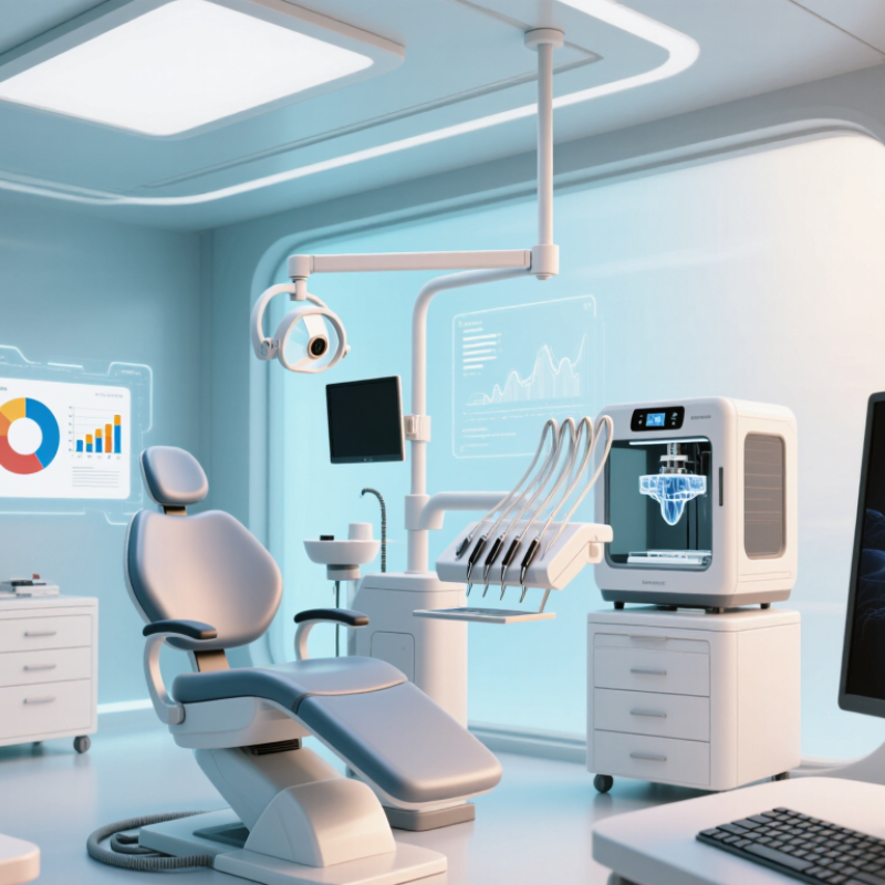
Some changes are visible. A printed surgical guide sits beside the treatment plan; a patient studies a scan on a chairside screen. Other changes are less obvious, such as software flagging an image for closer review. The distinction matters. These systems can support clinical decisions, but they cannot replace a full examination or a patient’s preferences. And new tools may introduce costs, workflow friction, or errors that deserve honest attention. This overview explores where emerging technologies may help, what questions clinicians should ask, and why thoughtful adoption—not novelty alone—will shape dentistry’s next chapter.
2026 Best Dental Technologies for the Future of Dentistry

Defining Dental Technology and Its Role in Dentistry in 2026

In 2026, dental technology includes the tools and digital workflows clinicians use to examine teeth, plan care, and explain findings. It can involve digital imaging, intraoral scanners, computer-guided planning, and software that organizes patient records. These tools do not replace clinical judgment. They can make details easier to review, but images still need careful interpretation. A bright screen is not proof of a complete diagnosis.

Tips: Ask what a scan or image shows, and how it affects your options. Keep copies of relevant records when changing providers. Clear explanations matter as much as new equipment.

The practical value depends on fit. A digital scan may capture the shape of a tooth without impression material, while planning software can help a clinician compare measurements before treatment. Yet equipment needs proper training, maintenance, and sensible use. If a scan is unclear, repeating it or choosing another assessment may be more responsible than trusting a neat-looking image. Technology can also add cost or complexity, and not every patient needs every tool. There is room for improvement: digital systems can feel efficient while still leaving patients unsure about what happens next. A good dental visit connects the data to a plain explanation, realistic choices, and follow-up care.
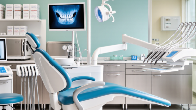

Digital Imaging and AI for Diagnosis and Treatment Planning

Digital imaging is making dental examinations more detailed and easier to compare over time. A small intraoral camera can show a chipped edge or irritated gum on a chairside screen. Digital radiographs reveal structures beneath the surface, while three-dimensional imaging may help assess complex anatomy when clinically appropriate. More detail is not automatically better. Each scan should answer a specific clinical question and use an appropriate level of exposure.

Artificial intelligence can help flag patterns in images, such as possible decay or changes near a tooth root. It can also support treatment planning by organizing measurements and comparing current images with earlier records. A dentist still needs to examine the patient, review symptoms, and interpret the findings. AI can miss subtle problems or highlight harmless variations. That can create extra worry.

The most useful workflow is collaborative: software points to an area, and the clinician checks it against the full picture. Patients should be able to see their images and hear, in plain language, what the findings may mean. Clear records also make follow-up comparisons more reliable. Yet image quality, software limits, and individual anatomy can complicate interpretation. That uncertainty deserves attention, not a confident-looking score.

2026 Best Dental Technologies for the Future of Dentistry — Digital Imaging and AI for Diagnosis and Treatment Planning
Technology Primary Clinical Use Typical Data or Output How AI Can Assist Key Advantages Important Considerations
Digital intraoral radiography Detecting and monitoring caries, periapical changes, and periodontal bone levels. Two-dimensional bitewing and periapical images. Can flag areas for clinician review, help measure findings, and support comparison with prior images. Images are available quickly and can be stored and compared digitally; exposure can be adjusted for the examination. Image quality depends on positioning, exposure, and interpretation. AI findings are aids, not definitive diagnoses.
Panoramic radiography Broad overview of the teeth, jaws, and surrounding structures; often used in assessment and referral planning. A single two-dimensional image of both dental arches and portions of the jaws. May help identify structures or regions that warrant closer clinical assessment. Provides a wide field of view in one examination. Superimposition and image distortion limit detail; it does not replace targeted intraoral imaging when indicated.
Cone-beam computed tomography (CBCT) Selected cases requiring three-dimensional assessment, such as implant planning, impacted teeth, or complex anatomy. Three-dimensional volume that can be viewed in multiple planes. May assist with segmentation, measurements, and highlighting anatomical structures. Shows spatial relationships that are not visible in conventional two-dimensional images. Uses ionizing radiation. Use should be justified for the individual patient, with the smallest suitable field of view and exposure settings.
Intraoral optical scanning Digital impressions for restorative, orthodontic, and appliance workflows. Three-dimensional surface model of teeth and nearby soft tissues. Can support automated measurements, align scans over time, and identify areas for review in selected workflows. Captures surface geometry without impression material and can be shared digitally with a laboratory. Records visible surfaces rather than internal tooth structures; accuracy can be affected by movement, moisture, and scan coverage.
AI-assisted image analysis Decision support for reviewing dental images and prioritizing areas for further assessment. Image annotations, probability scores, or highlighted regions, depending on the system. Algorithms analyze image patterns and present potential findings for clinician verification. May support consistency and help focus attention on areas that need review. Performance varies by task, image quality, and patient population. Outputs require professional review and should be interpreted with clinical findings.
Digital treatment-planning and CAD/CAM tools Planning restorations, orthodontic movement, and selected surgical or prosthetic procedures. Digital models, proposed designs, measurements, and simulated treatment stages. May automate measurements or generate draft designs that clinicians can edit. Supports visualization, communication, and transfer of digital records between clinical and laboratory workflows. Plans depend on accurate records and clinical judgment; a digital simulation is not a guarantee of the final result.
Longitudinal imaging and record comparison Monitoring changes in caries risk, bone levels, tooth position, or treatment progress over time. Side-by-side images, aligned scans, or measured differences between visits. Can help identify changes and bring potentially meaningful differences to the clinician’s attention. Provides a structured way to compare current records with earlier records. Comparisons are most useful when image technique and positioning are sufficiently consistent; differences should be confirmed clinically.

Advanced Tools for Restorative, Surgical, and Preventive Care

2026 Best Dental Technologies for the Future of Dentistry

Advanced Tools for Restorative, Surgical, and Preventive Care

Digital tools are changing how clinicians plan restorative, surgical, and preventive care. Intraoral scanners create detailed 3D impressions without traditional trays. Computer-aided design and manufacturing can help shape crowns with a precise fit. Still, a clean digital model does not guarantee a comfortable result. Clinicians need to check bite, margins, and patient comfort in person. Small adjustments matter.

For surgery, three-dimensional imaging can reveal bone structures that ordinary images may not show clearly. Guided planning helps clinicians map an approach before treatment, while real-time monitoring can support careful execution. These technologies can improve visibility and planning, but they cannot remove every risk. Image quality, clinician judgment, and the patient’s health all matter. A scan is useful, not infallible.

Preventive care also benefits from digital tools. Intraoral cameras can help patients see small areas of wear or plaque during an exam. Risk-assessment software may organize information such as medical history and past decay, but it should support—not replace—a clinician’s assessment.

Tips: Ask what a scan shows, how it changes your care plan, and whether follow-up is needed. Keep routine cleanings; technology works best alongside consistent care.

Connected Dental Workflows and Patient-Centered Treatment

Connected dental workflows make care feel less like a series of disconnected appointments. Digital scans, imaging, treatment plans, and follow-up reminders can travel together, helping clinicians explain what comes next. A patient can see a scan beside a simple care plan, rather than trying to remember a rushed conversation. A scan is not empathy, though. Teams still need to check understanding and invite questions.

The World Health Organization’s 2022 Global Oral Health Status Report estimates that oral diseases affect nearly 3.5 billion people worldwide. That scale makes access and continuity important. Connected tools may reduce repeated data entry and help teams spot missed follow-ups, but they do not guarantee better outcomes. The weak point is familiar: systems can connect while patients still repeat their story. Privacy, accurate records, and staff training matter as much as the software.

Tips: Before adopting a tool, map one patient journey, from booking to post-treatment check-in. Test whether information moves reliably between steps, and ask patients where instructions feel unclear. Small fixes count. Track missed appointments and follow-up completion, then review the results with the care team.

How to Evaluate Emerging Technologies for Clinical Use

2026 Best Dental Technologies for the Future of Dentistry
How to Evaluate Emerging Technologies for Clinical Use

A promising dental technology should solve a defined clinical problem, not merely produce a polished screen. Start by asking what decision it changes: earlier detection, clearer treatment planning, or fewer repeat visits. The WHO Global Oral Health Status Report (2022) estimates that 2 billion people have caries in permanent teeth, with 514 million children affected in primary teeth. The need is substantial, but need alone does not prove a tool works.

Check clinical evidence in patients resembling those in your practice. Look for prospective studies, independent validation, and clear reporting of false positives and missed disease. Test difficult cases, including restorations, mixed dentition, and low-quality images. Small gains matter. So do extra alerts that lead to unnecessary follow-up. A controlled pilot can compare the new tool with current care, tracking diagnostic changes, chair time, patient understanding, and cost.

Then examine safety, data handling, accessibility, and fit with existing workflows. Ask who reviews uncertain results and how errors are documented. Staff training takes time; a technically accurate tool may still disrupt a busy appointment. We should admit that early studies can look better than everyday use. Set a review date, define acceptable performance in advance, and pause adoption if real-world results fall short.